Congenital and Acquired Neuromuscular and Genetic Disorders
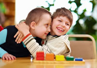
Cerebral Palsy
Cerebral Palsy is an umbrella term that describes damage to a child’s developing brain whilst either in the womb, during, or after birth. The way in which a child develops is affected, as the damage occurs in the part of the brain that deals with motor control, thereby affecting the child’s ability to control their movements and to hold postures.
 A diagnosis of Cerebral Palsy is usually made at around 12 to 18 months of age, providing that problems have been detected and an appropriate referral made to a Consultant specialising in this area. Some milder forms of Cerebral Palsy are harder to detect and difficulties may be picked up when developmental milestones are not being achieved, such as supported standing from sitting, or when a child appears ‘floppy’ or moves their limbs stiffly.
Every child with Cerebral Palsy is affected differently, as movement problems are specific to each child. Movement difficulties may be minor but can also be more profound, and some children may have other associated disabilities such as a learning disability, visual or perceptual problems, or epilepsy. These problems can affect the way in which your child interacts with their environment and the people around them, and ultimately affects their quality of life.
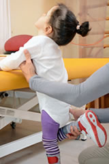
Physiotherapy intervention is important from an early stage following diagnosis, and a Specialist Paediatric Physiotherapist can work with a child with Cerebral Palsy, alongside their parents / carers and teachers, to help maximise their functional and emotional capacity, their growth and development, and to support a healthy and happy life.
Physiotherapy management and input varies with each child, as their needs and clinical presentation are unique to them. Movement difficulties and physical impairment vary, depending upon the type of Cerebral Palsy affecting the child. These can include Spastic Cerebral Palsy, Athetoid Cerebral Palsy and Ataxic Cerebral Palsy. Patterns of movement vary considerably between the three types and therefore, although the focus of physiotherapy is to facilitate and improve movement and function, its application and rehabilitation approaches must be specific to each individual child.
It is important to explain that Cerebral Palsy cannot be cured. Early detection and physiotherapy input are vital in order for your child to have the best functional outcome possible.
Children with Cerebral Palsy often require rehabilitation on a long-term basis, in order to promote function whilst they are growing, thereby supporting their development. Using hands-on therapeutic techniques, specific stretches and exercise programmes, a Specialist Physiotherapist can optimise a child’s muscle strength thus preventing further deterioration, and address issues with co-ordination / balance, abnormal movement patterns and sensory difficulties, and also manage secondary problems such as joint stiffness and soft tissue contractures. With regular and focused input, physiotherapy can help by increasing the potential to learn to walk, and by training motor and sensory processing skills that are necessary to support the child’s ability to function and interact as they grow older. In the longer term, it can help to develop independence and self-sufficiency in individuals affected by Cerebral Palsy.
Please get in touch if you feel that your child may be showing signs of movement difficulties or developmental delay. Our Specialist Team can assess your child and, if necessary, refer them onto an appropriate Specialist Consultant to confirm the diagnosis. Your child will be assessed through a process of observation and a hands-on examination, in order to identify their current functional capabilities and any difficulties. Following this, an individually-tailored management plan will be formulated and recommended in order to address their specific issues and needs.
We can provide specialist physiotherapy management, support and education for you and your child if they have recently been given a diagnosis of Cerebral Palsy, and we can also work in conjunction with any NHS physiotherapy rehabilitation your child may be receiving, in order to optimise the therapeutic outcome. Additionally, our Specialist Physiotherapy Team is experienced in providing rehabilitation for children who have received a botox injection, or undergone a surgical release of their tendons and tight soft tissues.
Selective Dorsal Rhizotomy
This neurosurgical procedure is performed on the lower spinal cord (usually between L2-S2) to treat spasticity in the lower limbs. It is major, irreversible surgery that requires intensive post-operative physiotherapy rehabilitation in order to maximise the potential gains of the surgery.
Surgically, the lower spinal nerves are selected and separated via electrical stimulation before certain nerve fibres are cut. The aim is to cut the sensory and motor nerve fibres that lead to muscle spasticity in individuals whose neurological condition causes reduced control over these spinal nerves, for example, children with certain presentations of Cerebral Palsy and other neurological disorders that cause spastic diplegia and quadriplegia.
It is important to mention that SDR is not a cure for conditions such as Cerebral Palsy, nor appropriate for all forms of Cerebral Palsy or all other neurological conditions. Some neurological conditions may have associated problems such as hip displacement or spinal scoliosis that may inhibit success of the SDR surgery, and therefore mean that these individuals are unsuitable for this procedure.
The surgery aims to relieve spasticity without losing the other necessary functions controlled by the spinal nerves, by carefully and precisely selecting the correct neural fibres during surgery. With reduced spasticity, the muscle groups can then begin to work more normally which then promotes more normal mobility and function. If spasticity is relieved, muscle scarring through contractures can be prevented and subsequently joint mobility is vastly improved.
Physiotherapy and occupational therapy begin on the 3rd day post-surgery, and are essential components of the entire process to ensure optimal results from the procedure. Physiotherapy is advised on an intensive basis, several times a week for 3 months to a year post-op, working closely with the individual and their family to facilitate normal muscle firing and to re-educate normal movement, walking and function.
Currently there is limited provision of this kind of surgery within the UK. Indeed most individuals who are suitable candidates for SDR travel to the USA for their surgery, and are self-funding with costs around £30-40,000 for the surgery and its follow-up care.
We are fortunate to have a highly specialist team of physiotherapists who are experienced in providing the extensive post-operative physiotherapy rehabilitation that is necessary for children following this procedure. Our team currently provides intensive post-operative rehabilitation for a number of children following SDR operations in the USA and in Leeds. Please feel free to get in touch if you would like to know more about this type of procedure and its aftercare.
Other useful links and resources regarding SDR are:
- https://www.scope.org.uk/advice-and-support/selective-dorsal-rhizotomy/
- ‘What’s the Evidence? Selective Dorsal Rhizotomy’, published by Cerebra
- ‘Treatment of Spasticity in Cerebral Palsy by Selective Dorsal Rhizotomy’, published by the National Institute for Health and Clinical Excellence (NICE)
- Email: info@yorkshirechildrensphysio.com
Erb’s Palsy
Erb’s Palsy is a type of birth injury to the brachial plexus. The brachial plexus is a group of nerve branches, bundled closely into the area between the neck and the shoulder, which ultimately provides movement and sensation to the upper limbs. Injury can occur during the delivery process, if a baby’s neck is overstretched to one side, the head is distracted from the neck, or excessive traction is placed through the shoulder, causing the brachial plexus on that side to be compromised.
When the brachial plexus is damaged during the birthing process, the upper limb on the affected side may present with weakness and/or altered sensation to varying degrees depending upon the severity of the injury. The C5 and C6 nerve branches are often affected, but occasionally also the lower cervical nerve branches.
The result may be an inability to lift the affected arm away from the side of the body (abduction) and the arm may hang next to the body, internally rotated from the shoulder joint, with the forearm also rotated inwards. There may also be some sensory loss and the loss of the biceps reflex. This movement paralysis has functional implications for a child who may, as a result of this type of injury, find it difficult to raise their arm above their head, dress themselves or feed themselves.
Some infants recover independently within their first few weeks of their life. Others may take longer (up to 2 years) and may require therapeutic intervention or exploratory surgery, whilst some never recover fully and may have long-term residual weakness and reduced overall muscle bulk of the affected arm. Recovery and prognosis tend to be more favourable if only the upper cervical nerves are affected. When both the upper and lower cervical nerves are affected, the presentation is more severe and may be termed ‘global / total brachial plexus palsy’.
If recovery is slow and little improvement is seen by the 3-month stage, a referral to a Consultant Neurosurgeon may be necessary. Physiotherapeutic intervention may include upper limb stretching and positioning (to prevent the development of contractures and to maintain joint range and soft tissue flexibility), massage therapy, active-assisted and passive movements of the arm (to stimulate normal movement patterns) and, in the older child, strengthening work and functional rehabilitation, if there are long-term impairments. Should surgical intervention be necessary, post-operative physiotherapy involvement would be similarly aimed at the restoration of movement, the prevention of soft tissue contractures, and the rehabilitation of strength and function in the growing child.
Parental involvement in the rehabilitation process is vital and is encouraged throughout all stages of the recovery period, to maximise the functional outcome for the child.
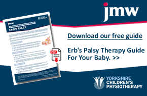
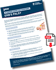 Download the Yorkshire Children's Physiotherapy Erb's Palsy Therapy Guide For Your Baby. Written by our very own Dr Paige Copeland in partnership with JMW Solicitors.
You can visit the JMW Solicitors Erb's Palsy page here.
The guide includes:
- Stretching exercises
- Strengthening exercises
- Sensory activities
Charcot-Marie-Tooth
Charcot-Marie-Tooth (CMT) is one of the most common hereditary neurological disorders affecting the peripheral nerves (i.e. those that lie outside the spinal cord and brain). The progression of this disease is gradual, affecting both sensory and motor nerves, therefore causing pain and altered strength, control and function of the limbs. Symptoms vary between individuals, and may become evident when a child begins catching their toes when walking, or losing their balance and falling frequently. The quality of their gait may initially be affected due to altered muscle control and sensation throughout the lower limbs, and they may have difficulty in grasping and holding objects, and opening bottles and jar lids.
As an individual with CMT grows and develops, the progressive weakening of their muscles can cause an imbalance to occur around their feet and ankles, leading to high arches, foot drop and/or curling of their toes (hammer toe). They may begin to report tingling or a burning sensation in their hands and feet and their fine motor skills may become increasingly difficult.
Physiotherapy for CMT should ideally begin as early in life as possible, at the highest point of muscle strength and function. The best results from rehabilitation are seen in individuals where the nerve degeneration is in the very early stages, and therefore the muscles are more responsive to a strengthening programme. Physiotherapy programmes may involve a mixture of strength training, soft tissue stretching, prevention of deformities, postural and core stability training, balance and co-ordination work, functional rehabilitation and exercise tolerance training to increase aerobic capacity, depending upon the specific needs and goals of the individual and the stage at which they present with their disorder.
Some individuals with CMT may require wrist supports, ankle braces, knee supports or foot orthoses (insoles) to help to control their joints and reduce deformity, to reduce pain and strain from their joints, and to enable them to continue with daily activities. Pain management may be an important facet of the overall management of CMT. Alternative therapies such as acupuncture and hydrotherapy may also be helpful in reducing muscular and joint pain.
Hereditary Spastic Paraplegia
Hereditary spastic paraplegia (HSP) is a hereditary neurological disorder, of which there are many types, which causes progressive spasticity and weakness of the muscles of the lower limbs, due to degeneration of the upper motor neurones in the brain and the spinal cord.
Symptoms may become evident at any age but commonly appear during a child’s teens or into their twenties and thirties. Initially, symptoms may be too mild to recognise as a disorder, for example an individual may begin to stumble more when walking, or find that they are tripping up and losing their balance more easily. As the lower limb dysfunction begins to worsen, movement difficulties become more obvious to the individual as they experience muscle spasms and their legs begin to feel weaker and stiffer. This progressive deterioration in the strength and function of the lower limbs means that individuals with HSP find it increasingly difficult to balance and walk as they get older.
Other common symptoms of HSP are altered bladder and bowel function, jumping feet (clonus), poor co-ordination of movement (secondary to muscle weakness and spasms), reduced sensation in the feet, and deformities of the feet such as high arches (pes cavus) due to muscle imbalance around the foot and ankle. There may also be other presenting medical conditions in those with HSP, some of which are unrelated.
The severity and rate at which the disease progresses varies between individuals, and therefore the rehabilitation required will be dependent upon the severity and stage of the disease. In the early stages, physiotherapy focuses on maintaining muscle strength, reducing spasm and preventing postural deterioration through core stability work and stretching. As the disease progresses, walking aids and functional adaptations may need to be introduced, with the key aims being to maintain a high quality of life, comfort and independence throughout the process.
Our Specialist Paediatric Physiotherapy Team will work closely with an individual with HSP to meet their specific needs and personal aims of rehabilitation, working alongside any other health professionals that may be involved, in order to optimise their functional capabilities and promote the physical and emotional well-being of the individual.
Muscular Dystrophy
Muscular Dystrophy is a lifelong hereditary (genetic) disorder of the muscles. Over time, the muscles become progressively weak, eventually breaking down and becoming replaced by fatty deposits. There are many forms of this condition, but the most common are Duchenne and Becker Muscular Dystrophy, the latter form being the less severe of the two.
 Problems often become apparent before a child is 5 years old. Initial symptoms such as weakness within the pelvic and shoulder muscles may be noticeable during periods of development and growth. For example, a child with this kind of muscular weakness may find it difficult to pull themself up into standing from sitting, or to get up from lying on the floor. Problems may also be evident when lifting a child up by holding them under their arms, as their shoulders may lift upwards (instead of being able to brace downwards) due to their muscle weakness.
As the child grows, the muscles of the body continue to progressively weaken, and in some cases, children find that by the age of 12 years, they need to use a wheelchair as their leg muscles have become too weak to support them. The muscles of the heart and chest also become weakened throughout the teenage years which may make breathing and tolerance of exercise difficult.
Physiotherapy intervention aims to maintain muscle capacity and mobility of the joints as far as possible, maximise function, and help to prevent deformities of the joints that have poor muscular support (such as within the spine). Walking aids and splints can also help to maintain a child’s ability to sit and stand for as long as possible. Physiotherapy also helps by providing a support network for families and carers involved in the management of a child with Muscular Dystrophy, and focuses on helping to give a child with this condition the best possible quality of life.
Microcephaly
Microcephaly is a condition that is present from birth, in which the baby’s head is much smaller than it should normally appear. The condition can result from foetal exposure to harmful substances, maternal diabetes, or may be as a result of an associated hereditary (genetic) condition. Acquired microcephaly can occur after birth if the baby’s brain becomes starved of oxygen or if an infection is present.
Symptoms vary with each child, but there are commonly problems with increased movement and spasticity of the limbs, and the child may have seizures, problems with developmental delay and mental retardation. Children with microcephaly may have a high-pitched cry and may also be poor at feeding.
Microcephaly is a lifelong disorder and some symptoms may become more evident as the child grows and ages. Physiotherapy management focuses on maximising functional ability through specific exercises, promoting normal development, and provides support to the child and their family, which continues through to their adulthood, to help improve their quality of life, confidence and their ability to integrate normally in society.
Spina Bifida
Spina Bifida is a condition caused by a fault in the way in which the spinal cord is formed whilst the baby is in the womb, which leads to an abnormal development of the spine. In this condition, a portion of the ‘neural tube’ of the embryo fails to join together at the end of the first month of foetal growth, leaving a gap in the spinal cord around which the spinal bones fail to develop. The presenting outcome depends upon the type of Spina Bifida, of which there are three:
Spina Bifida Occulta is the mildest form of Spina Bifida, and may even be present in individuals who have no symptoms or any awareness of the condition whatsoever. It is often detected as an incidental finding when an individual has an X-Ray of their lower back for an unrelated reason.
A baby or child with Spina Bifida Occulta may have a small patch of hair or a dimple in the skin over the level of the defect. Occasionally, there are related problems such as foot and ankle deformities (one or both sides of the body), knee flexion / hyperextension deformities, hip dislocation / dysfunction, pelvic tilt and leg length discrepancies, which may only be picked up later in life when these associated areas become symptomatic.
Mylomeningocele is a form of cystic Spina Bifida which can be serious in its presentation, as the spinal cord is usually damaged or poorly developed. Normal nerve function is affected below the damaged level of the spinal cord, causing paralysis and loss of sensation, and affecting the function of the bladder and the bowel. The extent of the impairment will depend upon the level at which the damage has occurred in the spine, and the degree of nerve damage at that level. Generally, lesions higher up the spine result in more severe paralysis and physical impairment.
A child born with this type of Spina Bifida may have other associated disorders such as hydrocephalus (fluid on the brain). This condition normally presents with a large mass bulging from the back (with or without skin coverage) and, in some cases, the spinal cord may actually be exposed.
Meningocele is another form of cystic Spina Bifida, but this one is less severe in its presentation than Mylomeningocele, with less spinal cord damage and therefore less neural impairment. An individual with this type of Spina Bifida may present with a small fluid-filled cyst on the outside of their back which is covered by a thin layer of skin. Meningocele is less commonly found however, than both Mylomeningocele and Spina Bifida Occulta.
Upon diagnosis of the latter two types of Spina Bifida, a child may be referred to a Paediatric Spinal Consultant or a Consultant Neurosurgeon, in order to assess the extent of their condition. In some cases, surgical repair and closure of the spinal cord lesion may be required.
Depending upon the level of neural impairment, a child with Spina Bifida may require specialist physiotherapy and occupational therapy intervention to address any difficulties and disabilities. Physiotherapy rehabilitation aims to optimise the use of the muscles and joints, prevent the formation of any secondary deformities and contractures, and maximise the child’s functional capabilities and independence. A Specialist Paediatric Physiotherapist can also advise on any aids and adaptations that a child whose normal function is significantly affected may require.
A strong support network is essential for families who are managing a child with a more severe form of this condition. Your physiotherapist can play a vital and supportive role, working alongside the family involved, and any other associated health professionals, helping to create an optimal lifestyle for both the child affected by Spina Bifida and the family as a whole.